Pelvic Floor Anatomy Ct

Mri Pelvis Anatomy Free Male Pelvis Axial Anatomy

The Pelvis Radiology Key
Http Pdf Posterng Netkey At Download Index Php Congress Ecr2014 Module Get Pdf By Id Poster Id 119484

Above Shows A Number Of Possible Measurements Using Mri Imaging A Download Scientific Diagram
Muscles Of The Pelvic Floor Anatomy And Function Kenhub
Ct Abdomen Pelvis Lower Axial Labeling Questions Radiology Case Radiopaedia Org

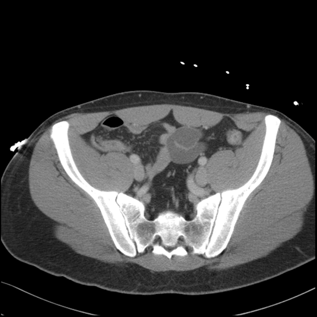
We created an anatomical atlas of abdominal and pelvic ct which is an interactive tool for studying the conventional anatomy of the normal structures based on a multidetector computed tomography.
Pelvic floor anatomy ct.

Pelvic Floor Muscles Pelvic Floor Muscles Ct

Eduardo D Campuzano Bs Rt R Mr Ct Ppt Video Online Download

Pelvic Floor Anatomy And Imaging Sciencedirect
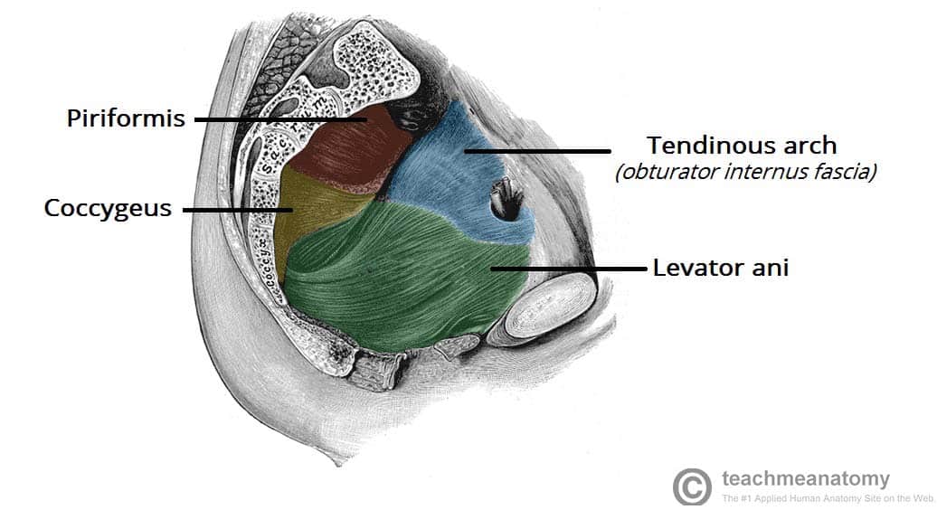
The Pelvic Floor Structure Function Muscles Teachmeanatomy
Https Www Abdominalradiology Org Resource Resmgr Education Dfp Pelvicfloor Anatomy Pelvic Floor Imaging And Ana Pdf
Https Www Ics Org Workshops Handoutfiles 000104 Pdf
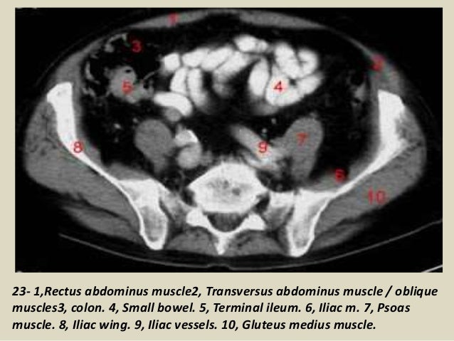
Presentation1 Pptx Ct Normal Anatomy Of The Abdomen And Pelvis

New Photos In Pelvis Anatomy
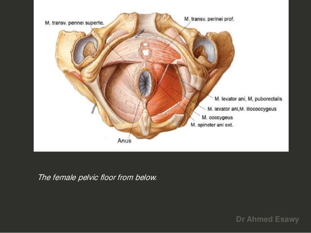
Anal Perianal Imaging Part 1 Ct Mri Anatomy Dr Ahmed Esawy

Pelvic Muscle Attachments A Classic Exam Question 1 Multiple Abdominal Muscles Iliac Crest Medical Anatomy Anatomy Medical
The Pelvis Ct Anatomy Mp4 Youtube Anatomy Pelvis Youtube

The Pelvis Anatomy Images Pelvic Floor Connective Tissues Bones
Muscles Of The Pelvis
Imaging Of Chronic Male Pelvic Pain What The Abdominal Imager Should Know Springerlink

Male Pelvis Sagittal Pelvis Anatomy Pelvic Floor Anatomy

Pelvic Diaphragm Of Male Superior View Www Anatomynote Com Pelvic Diaphragm Pelvis Anatomy Sacroiliac

Bulbospongiosus Origin Insertion Innervation Function Kenhub

Pelvic Floor Dysfunction And The Problem With Kegels Hands On
Https Encrypted Tbn0 Gstatic Com Images Q Tbn 3aand9gctpp0hdqwjnelsivrf64qutc9lu0ft7k Ajm10khrmneu9ez8uk Usqp Cau

Pin On Male Pelvic Pain

Pin By Pelvic Guru Llc On Pelvic Anatomy Pelvic Floor Pelvic Floor Exercises Floor Workouts

The Role Of Pelvic Floor Muscles In Male Sexual Dysfunction And Pelvic Pain Sexual Medicine Reviews
Https Pubs Rsna Org Doi Pdf 10 1148 Rg 345140137

Source : pinterest.com
