Mesothelial Cells In Pleural Fluid Tb Or Not Tb

Pdf Mesothelial Cells In Pleural Fluid Tb Or Not Tb

Pin By Diz F On Learning Chemistry Nurse Teaching Nursing Students Nurse

Tuberculous Pleural Effusion Shaw 2019 Respirology Wiley Online Library

Pleural Fluid Serial Analysis Reveals Lymphocytic Predominance Few Download Scientific Diagram
Http Www Smgebooks Com Tuberculosis Chapters Tb 18 20 Pdf

Accuracy Of Real Time Pcr Testing In Pleural Fluid For Tb Diagnosis Download Table
Eighty five samples of pleural fluid obtained from 76 patients with biopsy proven tuberculous pleurisy were examined cytologically.
Mesothelial cells in pleural fluid tb or not tb.
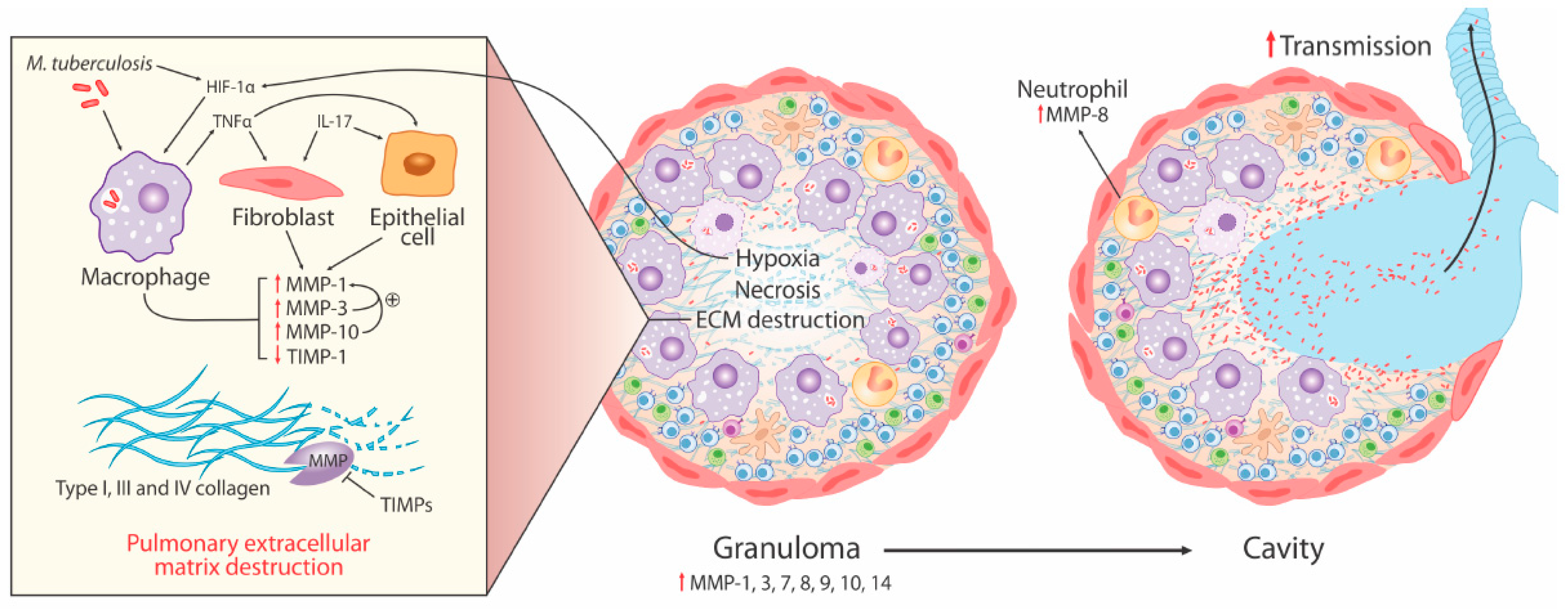
Ijms Free Full Text Matrix Metalloproteinases In Pulmonary And Central Nervous System Tuberculosis A Review Html
Were Sent For Cytological Histological And Microbiological Analysis Cytology Revealed Reactive Mesothelial Cells Histology Confirmed Endometrial Tissue With Abundant Well Formed Glandular Structures Confirming The Diagnosis

Tuberculous Pleural Effusion Respiratory Care
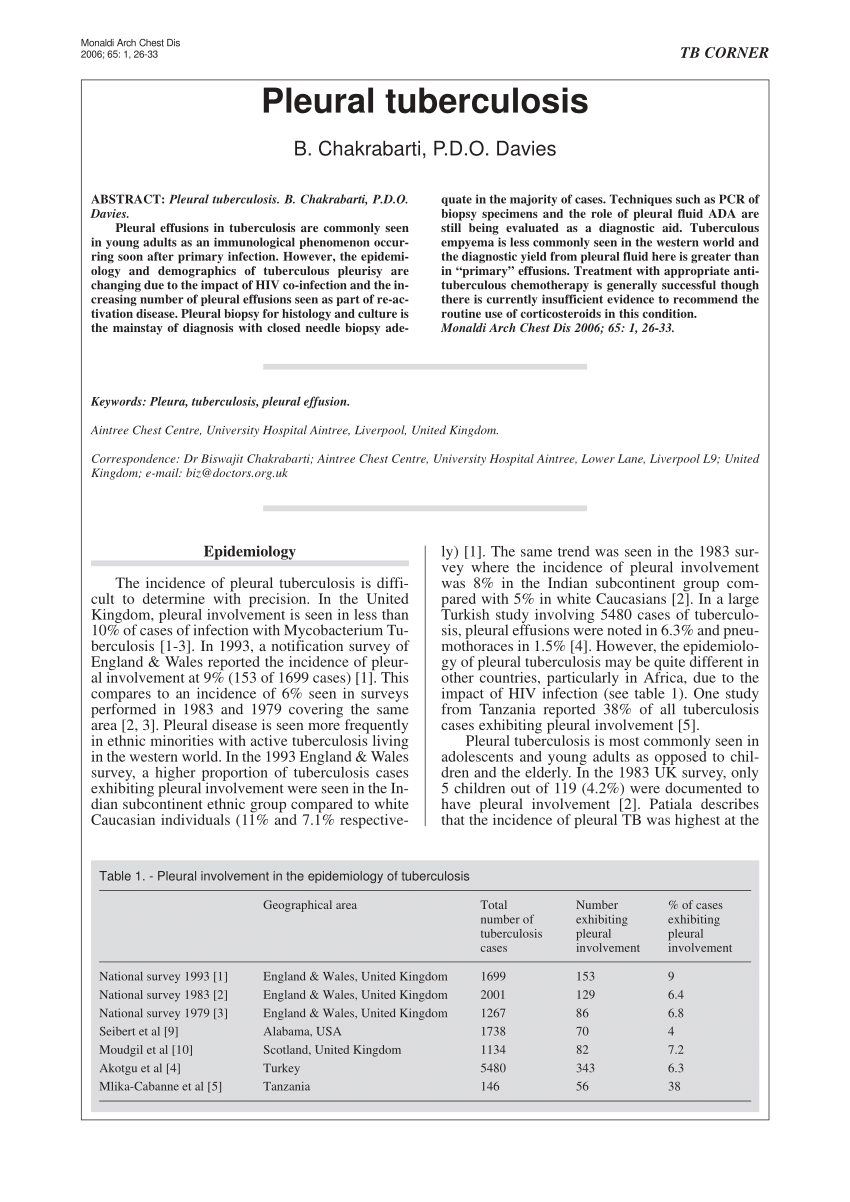
Pdf Pleural Tuberculosis
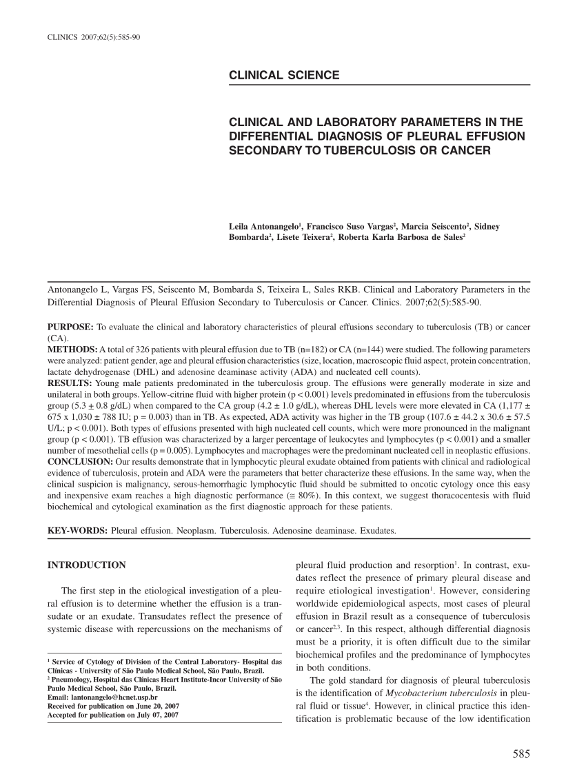
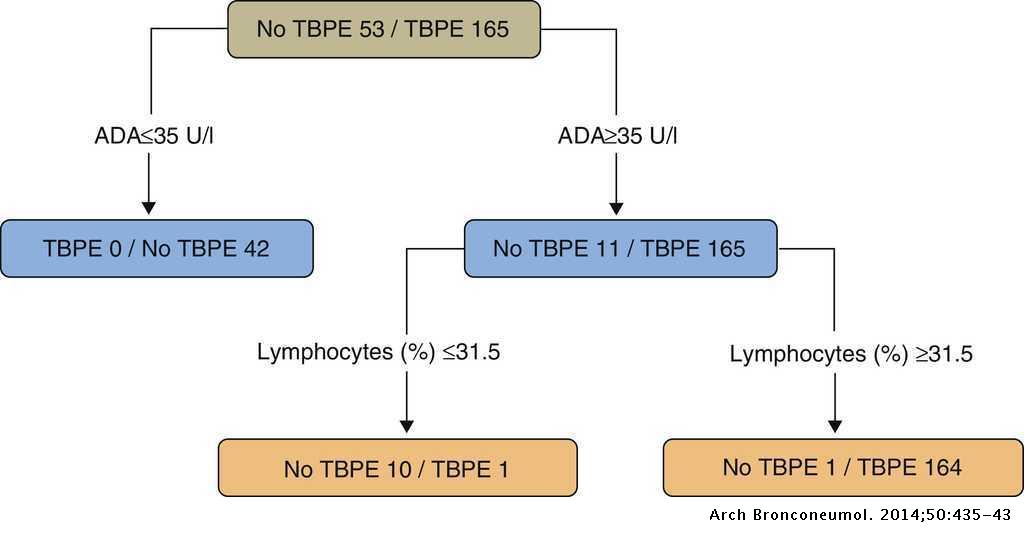
Pdf Clinical And Laboratory Parameters In The Differential Diagnosis Of Pleural Effusion Secondary To Tuberculosis Or Cancer

Tuberculosis Pleuritis Eurocytology
Https Jebmh Com Latest Articles 97110
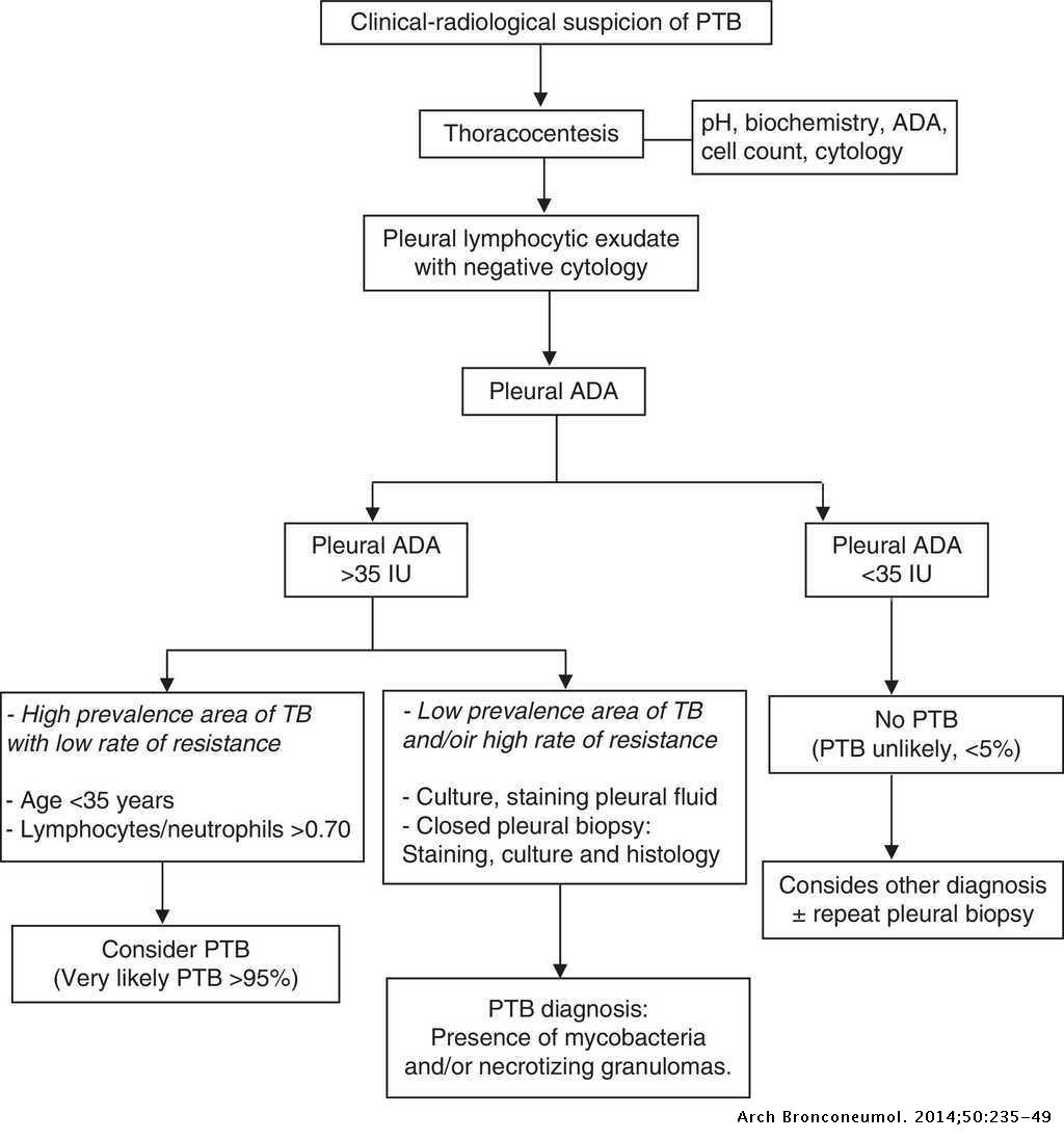
Recommendations Of Diagnosis And Treatment Of Pleural Effusion Update Archivos De Bronconeumologia
Tuberculous Pleural Effusion Archivos De Bronconeumologia

Flow Cytometry Comparison Between Peripheral Blood And Pleural Fluid Download Scientific Diagram
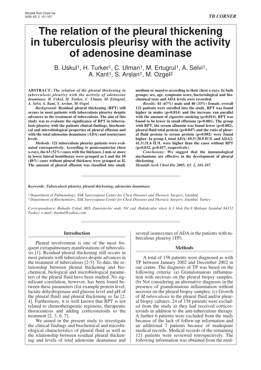
Pdf The Relation Of The Pleural Thickening In Tuberculosis Pleurisy With The Activity Of Adenosine Deaminase
Pleural Effusions That Can Be Diagnosed By Pfa Download Scientific Diagram
Pdf Diffuse Pleural Thickening Cases Of Pseudomesotheliomatous Adenocarcinoma And Pleural Tuberculosis
08 2019 Manila Difficult Pleural Management Pdf
Pdf Extrapulmonary Tuberculosis Semantic Scholar

Pleural Effusion Diagnosis Treatment And Management Abstract Europe Pmc
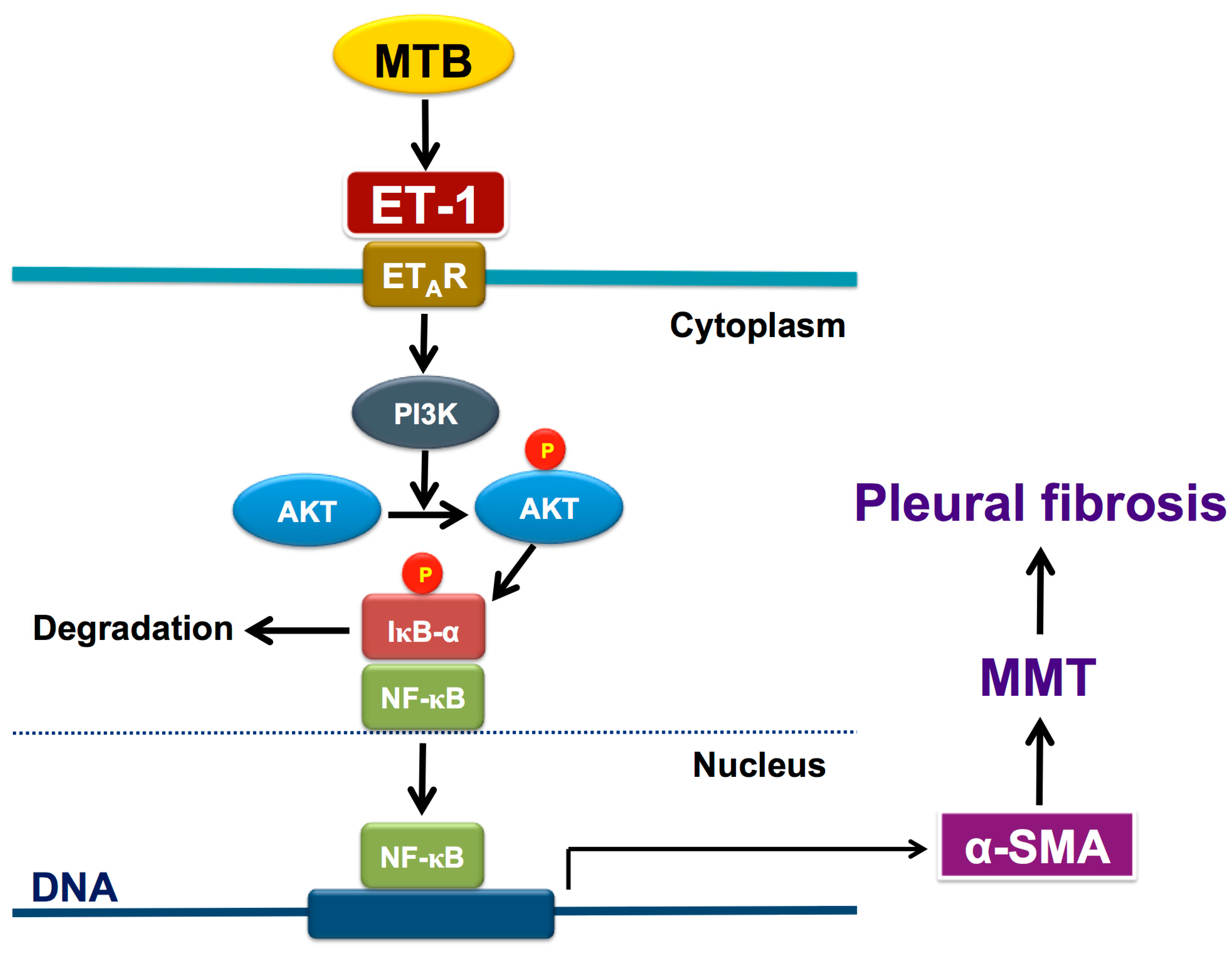
Jcm Free Full Text Endothelin 1 Induces Mesothelial Mesenchymal Transition And Correlates With Pleural Fibrosis In Tuberculous Pleural Effusions Html

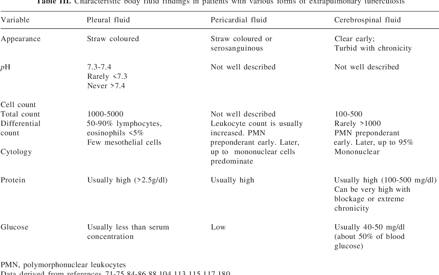
Biochemical And Cytological Characteristics In Tuberculous Pleural Download Table
Https Encrypted Tbn0 Gstatic Com Images Q Tbn 3aand9gcsoi9v0tyik1c1y6tlxd I66jv61b8fmdfeynwqjpvqc6llosxp Usqp Cau
Pdf Loculated Tuberculous Pleural Effusion Easily Identifiable And Clinically Useful Predictor Of Positive Mycobacterial Culture From Pleural Fluid
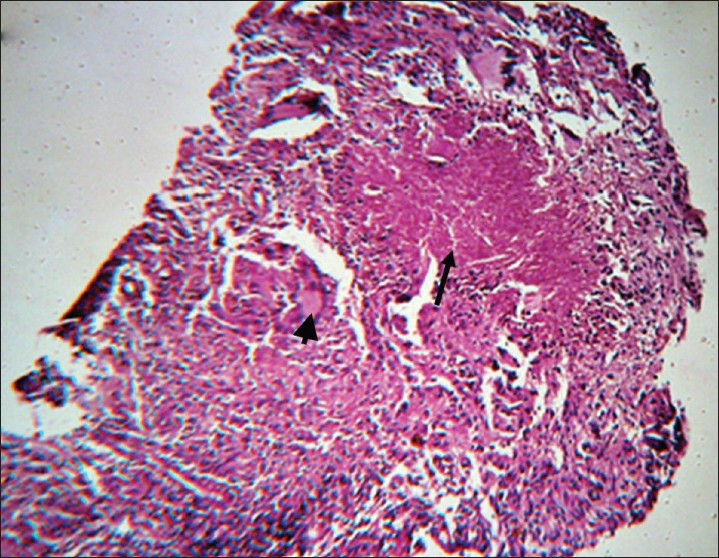
A Clinicopathological Study Of Tuberculous Pleural Effusion In A Tertiary Care Hospital
Pdf Ca 125 A Useful Marker To Distinguish Pulmonary Tuberculosis From Other Pulmonary Infections

Tuberculous Pleural Effusions Advances And Controversies Abstract Europe Pmc
Https Www Ghdonline Org Uploads Kohli Cohrane Xpert Eptb 2018 Nibrltx Pdf
Source : pinterest.com
